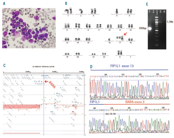
Figure 1.
Characterization of APL cells. (A) Morphology of the leukemia cells shows hypergranular promyelocytes with Auer roads in BM (100X). (B) A representative G-banded karyotype of the aberrant clone. The arrow indicates the derivative chromosome 17. (C) The panel shows the representative ideogram of the gain (40.8Mb) in chromosome 17q21.2q25.3, which results in the partial gain of the RARA locus and a possible rearrangement of this gene (arrow). (D) The sequence analysis of the identified fusion gene from the reverse sequence of RARA exon 3 identified FIP1L1 at exon 13 as the fusion partner gene. (E) Confirmation of the presence of the two reciprocal fusion transcripts by RT-PCR. Line 1: FIP1L1-RARA detection with a forward primer on FIP1L1 exon 10 (5′-ACAGCAGGGAAGAACTGGAA-3′), and a reverse primer on RARA exon 3 (5′-CCCCATAGTGGTAGCCTGAG). Line 3: RARA-FIP1L1 detection with a forward primer on RARA exon 1 (5′-ACACACCTGAGCAGCATCAC -3′), and a reverse primer on FIP1L1 exon 18 (5′-GTGTAGCTTCGGTGCTCTCC -3′). Lanes 2 and 4 are negative controls.

