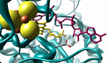
FIGURE 1.
Structure of the nucleotide-binding site of complex I with bound NADH. The protein is shown in blue, iron in red, sulfur in yellow, the FMN in yellow, and the bound NADH in magenta. The radius of the sulfur ion was set to 1.7 Å and that of the iron ions to 0.7 Å (48). The length of the hydrogen bonds between Glu-183F and the adenosine ribose hydroxyl groups are given in Å. The picture was drawn with Chimera (49) using the Protein Data Bank code 3IAM (13).

