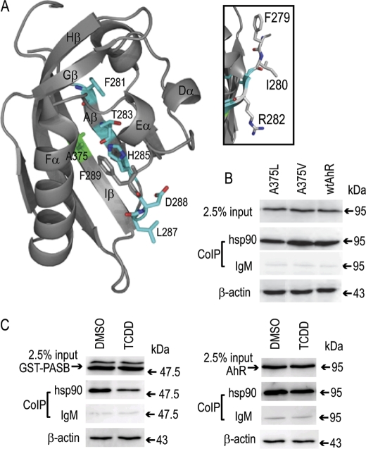
FIGURE 2.
The putative Hsp90-binding site spatially overlaps with the ligand-binding site in the PASB LBD. A, structure and labels are from the AhR PASB homology model (25). Internal Hsp90-interacting resides are shown in cyan, and Ala-375 (implicated in ligand binding) is shown in green. B, Hsp90 binding for mutant AhRs was carried out as described in the legend to Fig. 1B. C, TCDD treatment results in Hsp90 dissociation from the AhR PASB LBD. COS-1 cells transiently transfected with either the GST-PASB (left panel) or wtAhR (right panel) expression plasmid were incubated with TCDD (or solvent control) for 3 h, and cell lysates were subjected to co-immunoprecipitation (CoIP) assay as described in the legend to Fig. 1B. Western blot analysis was carried out using anti-PASB fragment antibody SE-8 for GST-PASB and anti-AhR antibody M20 for the wtAhR. Antibody SE-8 also detected an additional slower migrating nonspecific band (left panel), which did not bind Hsp90 and likely does not represent the functional AhR fragment. Results are representative of three independent experiments.

