FIGURE 1.
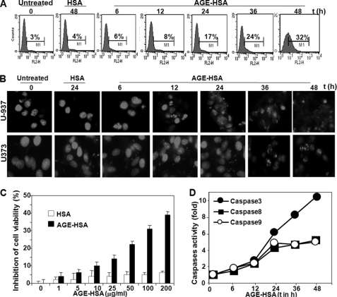
Effect of AGE-HSA on cell death. A, U-937 cells were treated with AGE-HSA (100 μg/ml) for different times. After these treatments, cell death was detected by annexin V-phycoerythrin and analyzed in FACS. U-937 cells were treated with AGE-HSA (100 μg/ml) for different times. Then cells were washed, fixed with methanol, and stained with propidium iodide. B, cells were then taken in slides and visualized in fluorescence microscopes. U-937 cells were treated with different concentrations of AGE-HSA in triplicate for 48 h. Cell viability was assayed using MTT dye. C, results are represented as inhibition of cell viability in percentage, which is calculated from mean absorbance ± S.D. of triplicate samples. D, U-937 cells were treated with AGE-HSA (100 μg/ml) for different times, and caspase 3, 8, and 9 activities were measured and indicated as -fold of activation considering untreated value as 1-fold.

