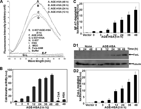
FIGURE 5.
Effect of AGE-HSA on intracellular Ca2+ release, calcineurin activation, NF-AT-dependent luciferase activity, and FasL expression. A, U-937 cells were treated with AGE-HSA (100 μg/ml) for different times. Intracellular free Ca2+ was measured using Fura-2AM as fluorescent probe in a fluorometer. Cells were treated with 100 μg/ml AGE-HSA for different times or CsA (2.5 μm) for 2 h and then treated with AGE-HSA for 48 h. B, calcineurin activity was assayed from whole cell extracts. U-937 cells were transfected with Qiagen SuperFect reagent for 3 h with plasmids for NF-AT or FasL promoter DNA that had been linked to luciferase (NF-AT-luciferase or FasL-luciferase) and GFP. After washing, cells were cultured for 12 h. The GFP-positive cells were, counted and transfection efficiency was calculated. C and D2, cells, treated with AGE-HSA (100 μg/ml) for different times, were extracted, and the luciferase activity was measured as per Promega protocol and indicated as -fold of activation. D1, the amount of FasL was measured from whole cell extracts upon similar treatment by Western blot. Error bars in B, C, and D2 indicate ± S.D. of triplicate samples.

