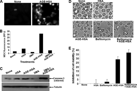
FIGURE 7.
Effect of AGE in inducing autophagy. A, U-937 cells were incubated with 100 μg/ml AGE-HSA for 48 h. Cells were washed and incubated with MDC (0.05 mm) for 10 min. The fluorescent cells were visualized under a fluorescence microscope. U-937 cells were pretreated with 3-methyl adenine (3MA, 5 mm) and then incubated with AGE-HSA (100 μg/ml) for 48 h. After incubation, cells were washed and incubated with MDC. B, intracellular MDC was measured by fluorescence photometry (excitation 380 nm and emission 525 nm). RFU, relative fluorescence units. C, the MDC incorporated was expressed as specific activity (arbitrary units). U-937 cells were pretreated with 3-methyl adenine (5 mm) and then incubated with AGE-HSA for 48 h. D, the cells were observed under bright field microscopy. U-937 cells, incubated with AGE-HSA (100 μg/ml) for 45 h, were co-incubated with bafilomycin (100 nm) for 3 h. E, cell viability was measured by MTT assay and indicated as inhibition of cell viability in percentage. Error bars in B and E indicate ± S.D. of triplicate samples.

