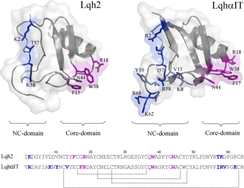
FIGURE 2.
Comparison of Lqh2 and LqhαIT. Top, bioactive surfaces of the two toxins. Lqh2 modeling was based on the reported structure of the almost identical toxin Aah2 (Protein Data Bank code 1AHO), and LqhαIT structure was determined (Protein Data Bank code 2ASC). The gray ribbons indicate the backbone structures covered by a semitransparent molecular surface of the toxins. Bioactive residues are shown as sticks (26, 29, 40). Residues of the core domain are colored magenta, and residues of the NC domain are colored blue. Bottom, sequence alignment of the two toxins. The bioactive surface of scorpion α-toxins consists of the conserved core domain (residues in magenta) and the diverse NC domain (residues in blue). Lqh2 and LqhαIT are similar in structure and share ∼70% sequence similarity, yet they exhibit opposing preferences for the mammalian brain and insect Navs (Table 1).

