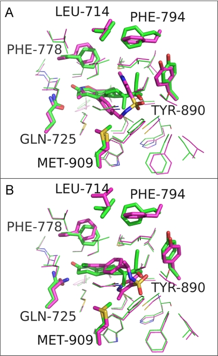
FIGURE 6.
A, overlaid binding pockets of PR-norethindrone complex (carbons colored green) with PR-OrgA complex (carbons colored magenta). Residues with the largest deviation in position between these two complexes are shown as sticks and labeled, and the remaining residues around the binding pocket are shown as lines. B, as per image A, but PR-OrgA complex replaced by PR-OrgB complex (carbons colored blue). Images were generated in PyMOL.

