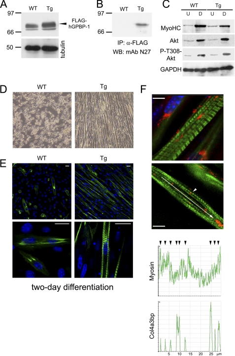
FIGURE 4.
Myoblast cell lines derived from Tg-hGPBP-1 mice show accelerated differentiation. A, lysates of undifferentiated myoblasts derived from wild type (WT) or Tg-hGPBP-1 (Tg) mice were analyzed by Western blot with specific antibodies for GPBP (mAb N27) or for tubulin, used as loading control. The arrowhead indicates the position of reactive polypeptide present only in Tg-hGPBP-1 lysates representing FLAG-tagged GPBP-1. B, the lysates in A were subjected to immunoprecipitation (IP) and Western blot analysis of precipitates (WB) using the indicated antibodies. Numbers and bars in A and B denote the size (kDa) and position of individual molecular mass markers. C, lysates (50 μg) of undifferentiated (U) and 4-day differentiated (D) WT or Tg-hGPBP-1 myoblasts were analyzed by Western blot using antibodies specific for the indicated proteins. D, the cells in A were differentiated for 2 days and analyzed by phase-contrast microscopy. Original magnification was ×100. E, representative confocal microscopy images of the cultures in D stained with antibodies for MyoHC visualization (green). Bars, 20 μm. F, representative confocal microscopy images of a differentiated Tg-hGPBP-1 culture stained with MyoHC-specific (green) and GPBP-specific (red) antibodies. The arrowhead denotes GPBP-1 intimately associated with an A band in a striated myofibril. Bars, 5 μm. The graphs represent the distribution of fluorescence intensity in the region indicated by the arrow in a nascent myofibril. The arrowheads denote co-localization of the indicated proteins.

