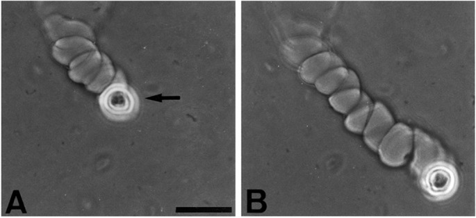
Figure 2.

Helical migration rate of a human keratinocyte. A human keratinocyte (strain YF29, culture V) was placed on a fibrin matrix as described in Fig. 1. It was photographed 8 h after plating (A) and 5 h later (B). Note the site of invasion of the matrix and the subsequent migration along a right-handed helix, whereas the helical axis remained nearly linear. By 13 h, the cell made 11 complete rotations around the helix axis and the helical path extended at a rate of 96 μm/hr for a total of 1,243 μm. Arrow points to the cell. (Phase contrast, bar = 50 μm.)
