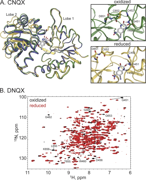
FIGURE 4.
A, crystal structures of the reduced (yellow, Protein Data Bank code 3T9V) and oxidized (green, Protein Data Bank code 3T9U) forms of the A452C/S652C GluA2 LBD mutant bound to CNQX. Also shown is the wild type structure bound to glutamate in blue. The disulfide bond is clearly formed in the oxidized case, with a dramatic change in the relative lobe orientations. B, the 1H,15N-HSQC spectra of the reduced (red) and oxidized (black) forms of the A452C/S652C GluA2 LBD mutant bound to DNQX. Specific chemical shift changes, consistent with the formation of the disulfide bond, are seen.

