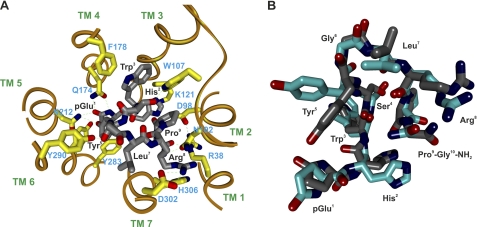
FIGURE 4.
Molecular model of the human GnRH receptor-GnRH I complex. A, A βII′-turn conformation of GnRH I derived from the NMR structure (PDB code 1YY1) was docked into the receptor model in the active conformation derived from the active state of bovine rhodopsin (3DQB) according to experimentally identified intermolecular interactions between GnRH I (gray) and the receptor contact sites (yellow). pGlu1 of GnRH I interacts with Asn212(5.39); His2 interacts with Asp98(2.61) and Lys121(3.32); Tyr5 interacts with Tyr290(6.58); Arg8 interacts with Asp302(7.32); and Pro9-Gly10NH2 interacts with Arg38(1.35) and Asn102(2.65). The H-bonds are indicated by dashed lines. The model suggests that the side chain of Gln174(4.60) makes H-bond contacts with pGlu1 of GnRH, whereas the aromatic ring of Phe178(4.64) interacts with the aromatic ring of Trp3 in GnRHs through displaced pi-stacking connections. B, NMR (cyan) and docked (gray) structures of GnRH I showing that the docked GnRH I has a trajectory similar to that of the NMR structure.

