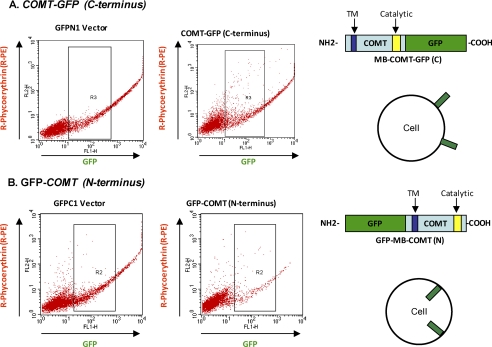
FIGURE 3.
Catalytic domain at the C terminus of MB-COMT is in the extracellular space revealed by flow cytometry. A, lymphoblastoid cells transfected with COMT-GFP (C terminus) and its GFPN1 vector were immunostained with R-PE-conjugated anti-GFP antibody and analyzed with flow cytometry. The strong R-PE signal from the COMT-GFP transfected cells indicates that the GFP tag is located in the extracellular space as illustrated by the depiction on the right. B, lymphoblastoid cells transfected with GFP-COMT (N terminus) and its GFPC1 vector were immunostained with R-PE-conjugated anti-GFP antibody and analyzed with flow cytometry. The R-PE signal from the COMT-GFP transfected cells is the same as that from the GFPC1 vector-transfected cells, suggesting that the GFP tag is located in the cytoplasm as illustrated by the depiction on the right.

