FIGURE 3.
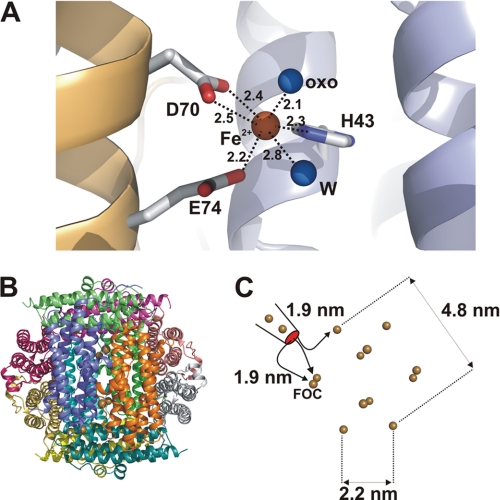
Structure of FOC and distribution of FOCs in dodecameric complex. A, structure of the FOC with the iron atom in brown and the ligating molecules (water (W) and the oxo atom (oxo)) in blue (bond lengths are given in Å). The iron atom has a pseudohexameric coordination sphere with two residues contributing three bonds (Asp-70 and Glu-74) from one subunit (in brown) and a third residue (His-43) located on the second subunit (marked in blue). B, dodecameric arrangement of AAH with all subunits color-coded by different colors. Small brown dots represent the positions of iron atoms bound in the AAHL structure, all of which are bound at the FOCs. C, schematic representation of the 12 iron atoms localized at FOCs and distances given in nm between these atoms. Distances between the entry of iron into the inner shell and the three nearest neighboring FOCs are marked.

