FIGURE 7.
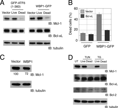
Analysis of Bcl-2 family proteins in apoptotic cells. A, specific reduction of Mcl-1 expression in apoptotic cells transfected with active ATF6 or WBP1. B, co-expression of Bcl-xL suppressed apoptosis induced by WBP1. C2C12 cells were grown in six-well plates and transiently co-transfected with GFP or WBP1-GFP (0.3 μg) and pcDNA3.1 vector or Bcl-xL (1.8 μg). C, down-regulation of Mcl-1 in cells that overexpressed Bcl-xL. C2C12 cells were electroporated with WBP1-GFP and Bcl-xL at a 1:1 ratio (w/w). Mcl-1 was analyzed by Western blot. The band intensity was normalized with α-tubulin as a standard. Representative data from three independent experiments are shown. D, specific reduction of Mcl-1 in apoptotic cells treated with ER stressors tunicamycin (TUN) and thapsigargin (TG). UT, untreated; IB, immunoblot.

