FIGURE 5.
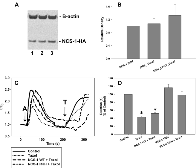
Overexpression of NCS-1 I35H rescues NCS-1 levels and the calcium signal in SHSY-5Y cells. A, representative Western blot shows that NCS-1 I35H-HA levels are not significantly changed with paclitaxel treatment. Lane 1, NCS-1 I35H-HA; lane 2, NCS-1 I35H-HA + paclitaxel; lane 3, NCS-1 I35H-HA + CAST + paclitaxel. B, NCS-1 I35H-HA was not decreased by paclitaxel treatment, nor was it enhanced by the addition of CAST prior to paclitaxel treatment (n = 3). *, p ≤ 0.05. All treatments were normalized to cells transfected with HA-tagged NCS-1 I35H. C, the intracellular calcium signal was reduced after a 6-h treatment with paclitaxel. The overexpression of NCS-1 WT prior to paclitaxel treatment was unable to rescue the calcium signal fully to that of control cells. However, cells expressing NCS-1 I35H prior to treatment with paclitaxel had calcium signals similar to that of control cells (15–20 cells, n = 3). (A, 1 μm ATP; T, 10 μm thapsigargin). D, comparison of the normalized response durations to 1 μm ATP in control cells, cells treated with paclitaxel, and cells overexpressing NCS-1 WT or I35H is shown. All treatments are normalized to control cells. All values are mean ± S.E. (error bars).

