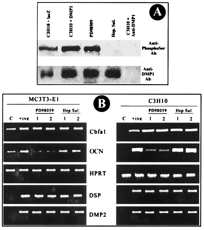
Figure 7.

Signaling pathway in DSP/DMP2 expression. (A) DMP1 phosphorylation. C3H10T1/2 cells were infected with adenoviral constructs. Total protein extracts were prepared as described in Materials and Methods and immunoprecipitated with affinity-purified anti-DMP1 antibody. (Top) Western blot against anti-phosphoserine antibody on DMP1 immunoprecipitates. (Bottom) Western blot performed with anti-DMP1 antibody on the DMP1 immunoprecipitates. (B) DSP and DMP2 expression. Cells (2 × 10⁶ cells per milliliter) were seeded 24 h before infection with Ad5/CMV-DMP1. Infection was carried out for 3 h, and fresh medium was added to the cells. A MAPK inhibitor (PD98059, 100 μg/ml) and a casein kinase II inhibitor (heparan sulfate, 100 μg/ml) were added along with fresh medium and incubated for another 24 h. Total RNA was isolated from cells, and RT-PCR was performed. C, Control cells infected with β-gal expression cassette. +, Positive control used for the experiment [infected with Ad5/CMV-DMP1 (sense) without inhibitors]. 1 and 2, Duplicates for the experiment. RT-PCR analysis for Cbfa1, OCN, DSP, and DMP2 was done with hypoxanthine phosphoribosyltransferase as a control.
