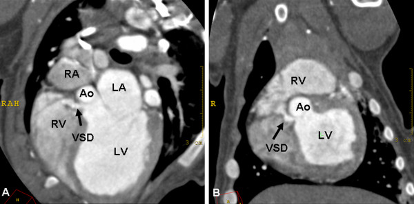
Figure 5.
Ventricular septal defect in a dog. Oblique transversal (A) and dorsal (B) MPRs of contrast-enhanced ECG-gated cardiac MDCT images. The images show the ventricular septal defect (VSD) as a small contrast-enhanced connection between the right ventricle (RV) and the left ventricle (LV). LA = left atrium, RA = right atrium, Ao = aorta.

