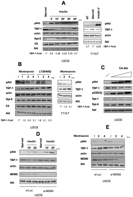
Figure 6. TBP-1 is a downstream target of Akt activation.
A: U2OS cells or T11hT cells were starved for 4 hrs and then treated with 10 ng/ml insulin for the times indicated. Activation of Akt/PKB was evaluated by Western Blot on whole protein lysates probed with anti-Phospho-Akt Ser473 and anti-Akt. Levels of endogenous TBP-1 and of two proteasome components (C8 and Rpt-6) were analyzed where indicated. TBP-1 bands intensity was measured by ImageQuant analysis on two different expositions to assure the linearity of each acquisition, each normalised for the respective actin values. Asterisk, fold value is expressed relative to the reference point, (i.e. TBP-1 levels in starved cells) arbitrarily set to 1. Representative of three independent experiments. B: U2OS cells or T11hT cells were treated, 24 hrs after plating, either with DMSO (/) or with 200 nM Wortmannin or 50 mM LY294002 for the times indicated. Cells were then lysed and Western Blot analysis was performed by using specific antibodies against Phospho-Akt Ser473, anti-Akt, anti-TBP-1, anti-C8 and anti-Rpt-6. TBP-1 bands intensity was calculated as in A. Representative of three independent experiments. C: U2OS cells were transfected with empty vector (lane 1) or increasing amounts of the constitutive active mutant of the Akt kinase (CA-Akt). After 24 hrs cells were lysed and whole cell lysates probed with anti-Phospho-Akt Ser473, anti-Akt, anti-TBP-1, anti-Rpt-1, anti-Rpt-6, and anti-phospho-GSK3b. D: U2OS cells were transfected with a siRNA directed against MDM2 or Luciferase, as control, at the final concentration of 10 nM. After 24 hrs, cells were starved for 4 hrs and then treated with 10 ng/ml insulin for the times indicated. Cells were then lysed and Western Blot analysis was performed by using specific antibodies against Phospho-Akt Ser473, TBP-1, MDM2, Akt, and actin. E: U2OS cells were transfected with a siRNA directed against MDM2 or Luciferase, as control. After 48 hrs, either DMSO (/) or 200 nM Wortmannin was added to the cells and left for the times indicated. Cells were then lysed and Western Blot analysis was performed by using specific antibodies against Phospho-Akt Ser473, TBP-1, MDM2, Akt and actin.

