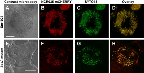
Figure 2. The S. meliloti bacA mutant is challenged with NCR peptides.
Microscopy of transgenic M. truncatula nodule cells expressing NCR035-mCHERRY under the control of the NCR035 promoter infected with either the S. meliloti wild-type (A–D) or BacA-deficient mutant (E–H) strains. Differential Interference Contrast microscopy (A,E), confocal microscopy of NCR035-mCHERRY localization (red) (B and F), bacterial localization with the DNA stain SYTO13 (green) (C,G), and an overlay of NCR035-mCHERRY and SYTO13 localization (D,H). In both the wild-type and BacA-deficient mutant infected nodules, NCR035 co-localizes with the bacteria. Scale bars are 10 µm.

