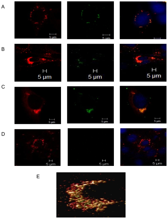
Figure 3. DENV-2 E protein traffics within the endosomes post internalization.
BHK-21 cells were infected at MOI of 5. Cells were stained for endosomes by adding lysotracker red to the cells 2 h prior to fixation. The cells were stained for DENV-2 E protein using mouse anti-E MAb followed by goat anti-mouse IgG FITC. Split images show DENV-2 E protein associating with endosomes at A] 0 min B] 30 min C] 2 h and D] 4 h. E] 3D reconstruction of the z stacked (thickness 3.30 µm) deconvolved image for the 30 min time point.

