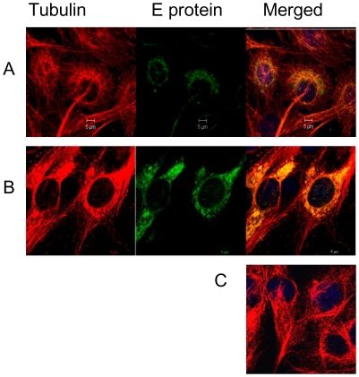
Figure 4. DENV-2 E protein traffics on microtubules.
BHK-21 cells infected at 1 MOI were fixed at different time points and stained for DENV-2 E protein and anti-alpha tubulin. The E protein was labelled with mouse anti-E MAb followed by goat anti-mouse IgG FITC conjugate. Cells were saturated with unlabeled goat anti-mouse IgG and then stained with mouse anti-alpha tubulin MAb followed by rabbit anti-mouse IgG TRITC. Infected cells showing colocalization of DENV E protein and alpha-tubulin, A] at 8 h and B] at 24 h. C] Mock infected cells showing distribution of alpha-tubulin.

