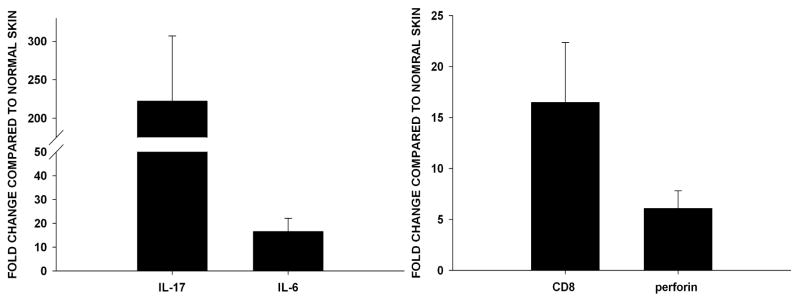
Figure 7. SC tumor sites express IL-17, IL-6, CD8 and perforin.
To determine the expression of IL-17, IL-6, CD8 and perforin in SC tumors Ad5E1 clone 2.1 tumor cells (5 × 10⁴) were injected in the skin of IFN-γ KO mice. After 7 days, skin samples were excised and RNA was isolated. The same qPCR procedure was done as described above. mRNA expression of (A) IL-17 and IL-6, (B) CD8 and perforin was observed in skin samples injected with tumor cells. Samples were normalized to GAPDH and compared to normal (naïve) skin (fold change +/− SE). Data are the combined results of eight individual mice. P values < 0.05 were considered significant as determined by Student’s t test.

