Fig. 4.
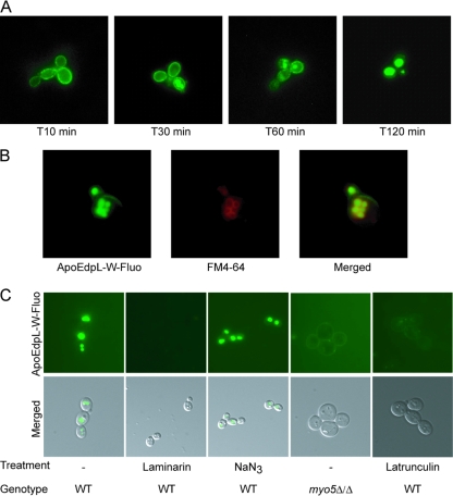
ApoEdpL-W is targeted to vacuoles through endocytosis. (A) Kinetic localization of ApoEdpL-W-Fluo in C. albicans SC5314 cells exposed to 5 μM ApoEdpL-W-Fluo. Images are representative of labeling distribution at each time point. (B) C. albicans SC5314 cells exposed to ApoEdpL-W-Fluo (40 μM) and stained with FM4-64. (C) C. albicans MYO5/MYO5 or myo5Δ/myo5Δ cells exposed to ApoEdpL-W-Fluo (5 or 20 μM) for 30 to 60 min. In the laminarin panel, ApoEdpL-W-Fluo was preincubated with 5 mg/ml laminarin for 1 h prior to being applied to cells. In the NaN3 panel, cells were incubated with 10 mM NaN3 for 1 h prior to being exposed to ApoEdpL-W-Fluo. In the latrunculin A panel, cells were incubated with 50 μM latrunculin A for 1 h prior to being exposed to ApoEdpL-W-Fluo.

