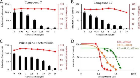
Fig. 2.
In vitro inhibition of hepatic Plasmodium berghei infection by compounds 7 and 10. (A to C) Compounds were added to Huh7 hepatoma cells 1 h before infection with luciferase-expressing sporozoites. An amount of DMSO equivalent to that in the highest compound concentration tested was used as a control. Forty-eight hours after the addition of P. berghei sporozoites, cell confluence (red lines on bar plots) was assessed by alamarBlue fluorescence, and the infection rate (bars) was measured by quantifying the luciferase activity by luminescence. The effects of different concentrations of compound 7 (A), compound 10 (B), and a 1:1 mixture of primaquine and artemisinin (C) are shown. Results are expressed as means ± standard deviations. (D) For each compound, the IC50 was calculated by sigmoidal fitting. Red, compound 7; orange, compound 10; green, primaquine plus artemisinin.

