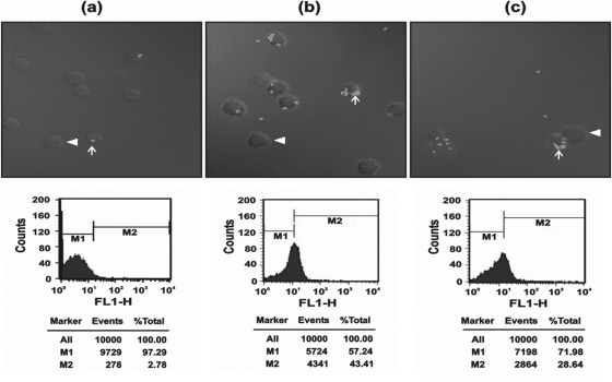
Fig. 10.
(a) Laser scanning confocal images of macrophage phagocytosis. (b) Medium control showing very few internalized FITC-labeled E. coli cells within the macrophages (×63 magnification). When macrophages were induced with LPS, phagocytosis was significantly increased compared to that of the scrambled peptide. (c) In LPS-induced macrophages treated with RVFHbαP (70.45 μM), the number of E. coli cells internalized within macrophages was significantly inhibited. Also shown are positions of plasma membranes (arrowheads) and labeled bacteria internalized within the macrophages (arrows). The images are representative of one of three identical experiments performed on three different days.

