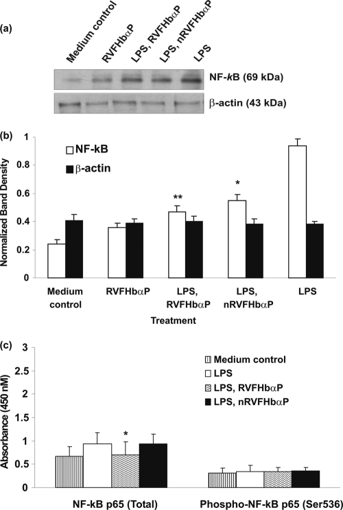
Fig. 9.
Western blot analysis of NF-κB expression in HeLa hVECs. Cells were seeded at a density of 106/well in 24-well plates and treated with LPS (10 μg/ml for 6 h), or LPS-induced (10 μg/ml for 6 h) cells were treated with RVFHbαP (70.45 μM for 1 h) or scrambled peptide (70.45 μM for 1 h). At the end of treatment, cells were collected and lysates were prepared and analyzed for NF-κB and β-actin expression by Western blotting as detailed in Materials and Methods. Expression of NF-κB was upregulated in LPS-induced cells and significantly attenuated following the treatment of LPS-induced cells with RVFHbαP in respect to the scrambled peptide. Values were calculated as the means ± SD of triplicate determinations and are representative of at least three separate experiments performed on different days. Representative image of Western blot analysis of NF-κB expression is shown. β-Actin blot confirmed roughly equivalent loading of protein samples. (b) A quantitative assessment of the intensity of each band was determined by densitometry. (c) Protein ELISA was carried out to confirm the data obtained by Western blot analysis, and the results are in agreement with Western blot results. Levels of significance (*, P < 0.05; **, P < 0.001; compared with the LPS-induced group) were calculated by an ANOVA test followed by a Bonferroni analysis.

