Fig. 2.
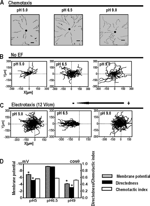
Extracellular pH plays different roles in chemotaxis and electrotaxis in Dictyostelium cells. (A) cAMP gradients were formed from the tip of a micropipette filled with 10 μM cAMP. Trajectories of cell migration toward cAMP in DB with different pH values are indicated. Dark spots represent the position of the micropipette. Scale bar, 20 μm. (B) Cell migration in random directions under control conditions without an EF although cells were bathed in DB with different pH values as indicated. (C) Cell migration trajectories in which the start point of each cell is set as the origin. Cells migrated cathodally in a pH of 6.5 in an EF. However, directed cell migration was significantly impaired under acidic (pH 5.0) or alkaline (pH 9.0) conditions. (D) The effects of extracellular pH on electrotaxis in Dictyostelium cells correlate with the effects on Vm. Dictyostelium cells bathed in pH 6.5 showed a greater Vm than that of the cells in pH 5.0 or pH 9.0 and significantly better electrotaxis. The changes in pH and corresponding changes in Vm did not significantly affect the chemotaxis (chemotactic index). *, P < 0.001 compared to that in pH 6.5.

