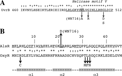
Fig. 5.
(A) Deduced amino acid sequence of the B. subtilis UvrB protein. Helicase motif V (12) is underlined and labeled. Mutations altering UvrB helicase and incision activity (G502R, G509D, and G509S) (25) are indicated by downward arrows. Valine 498 is boxed, and the V498A mutation in strain WN716 is indicated. (B) Deduced N-terminal amino acid sequences of AlsR and OxyR. The underlined amino acids are highly conserved among LysR family proteins (53). Alanine 21 in AlsR is boxed, and the A21D mutation in AlsR is indicated by the upward arrow. The downward arrows denote mutations in the OxyR HTH region leading to decreased DNA binding (16). The bottom line indicates the position and extent of alpha-helices (α1, α2, and α3) in the LysR HTH region (53). In both panels, the asterisks and colons denote identical and conserved amino acids. See the text for details.

