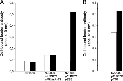
Fig. 3.
Cell surface location of the N terminus of the fusion peptide construct. Whole-cell ELISA on L. lactis NZ9000 cells for detection of surface-displayed prenisin anchor fusion protein (A) and angiotensin-(1-7) anchor fusion protein (B). Rabbit anti-nisin leader antibodies were allowed to bind to lactococcal cells displaying TB5 or TB2. Alkaline phosphatase-conjugated goat anti-rabbit IgG was added, and a color was generated by the addition of p-nitrophenylphosphate. White bars, uninduced cells; black bars, induced cells. A typical experiment is depicted. The experiment was repeated in more than three independent replicates with differences of ≤15%.

