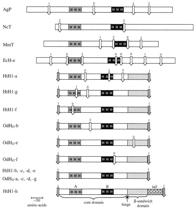
Figure 2.
Comparison of intron locations in molluscan hemocyanins and several related proteins. Internal introns are shown by white arrows, linker introns by gray arrows. The copper-binding sites (A and B), with their histidines (H) are also schematically included. The β-sandwich domain in molluscan FUs is indicated by the dotted region, with the hinge location marked. The unique tail of FU-h is also indicated. Notation (aside from molluscan hemocyanin FUs): AgP Anopheles gambia (insect) prophenoloxidase; NcT, Neurospora crassa (fungus) tyrosinase; MmT, Mus musculus (mouse) tyrosinase, EcH-e, Eurypelma californicum (tarantula) hemocyanin subunit e.

