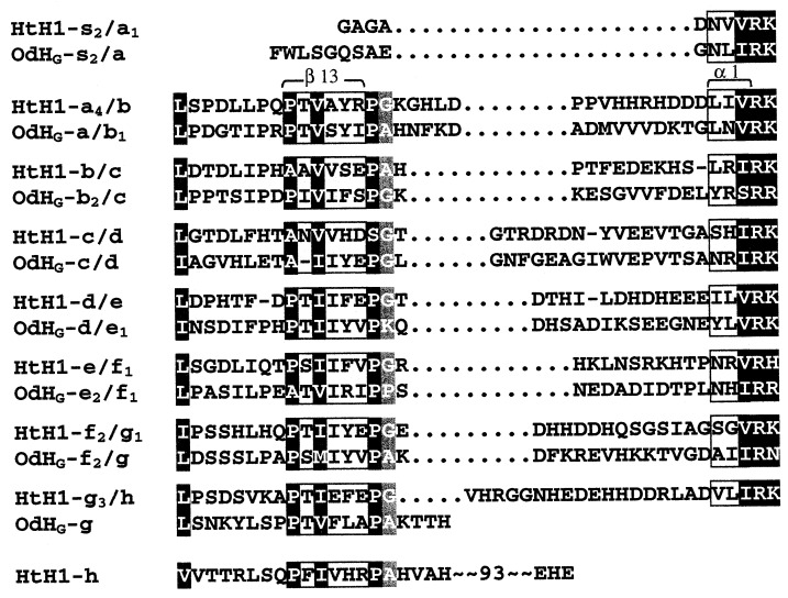
Figure 3.
Sequence surrounding the linker introns. The protein linker regions between the various FUs (designated by the respective exon pair) have been arranged so as to (i) align conserved residues upstream from the linker intron insertion sites (indicated by dotted lines) and (ii) align the (I/V)RK consensus downstream from the insertion site. Strongly conserved residues are marked in black; there are many more of them in the β-sheet region of each preceding exon, to the left of the intron insertion site. The residues to the right of the site constitute the protein linker sequence; there is little conservation here until the (I/V)RK motif, which marks the beginning of the first α-helix in the next exon. To make both alignments, it has been necessary to introduce gaps in some of the linker sequences; their positions are not well defined. The positions of two secondary structure elements as deduced from OdH-g (3) are marked in light gray, and the positional conservation of a small amino acid is marked in dark gray.

