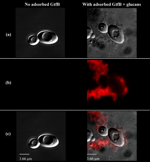
Fig. 2.
Visualization of glucans synthesized in situ by GtfB adsorbed to C. albicans SC5314 yeast cells. C. albicans cells were incubated with GtfB (or buffer) and washed to remove unbound GtfB. Cells with adsorbed GtfB (or without GtfB bound, as a control) were then exposed to sucrose, and glucan formation was assayed after 2 h of incubation. (a) DIC image of C. albicans cells after incubation with sucrose (100× oil objective, numerical aperture 1.4). (b) Image obtained with laser excitation at 633 nm for detection of glucans (Alexa Fluor 647). (c) Overlaid DIC and fluorescence images.

