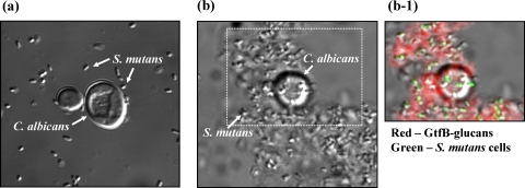
Fig. 3.
S. mutans UA159 cells binding to C. albicans SC5314 cells with and without surface-formed GtfB glucans. Cells of C. albicans (with or without surface-formed GtfB glucans) and S. mutans were incubated together (1 h) and then analyzed using DIC and fluorescence imaging. (a) DIC image of C. albicans and S. mutans cells after incubation (100× oil objective, numerical aperture 1.4). (b) DIC image of GtfB glucan on C. albicans and S. mutans cells after incubation. (b-1) Overlaid DIC and fluorescence images of the field of view selected in panel b. Laser excitation at 488 nm was used for detection of SYTO 9-labeled S. mutans cells (green), and laser excitation at 633 nm was used for detection of Alexa Fluor 647-labeled GtfB glucans (red).

