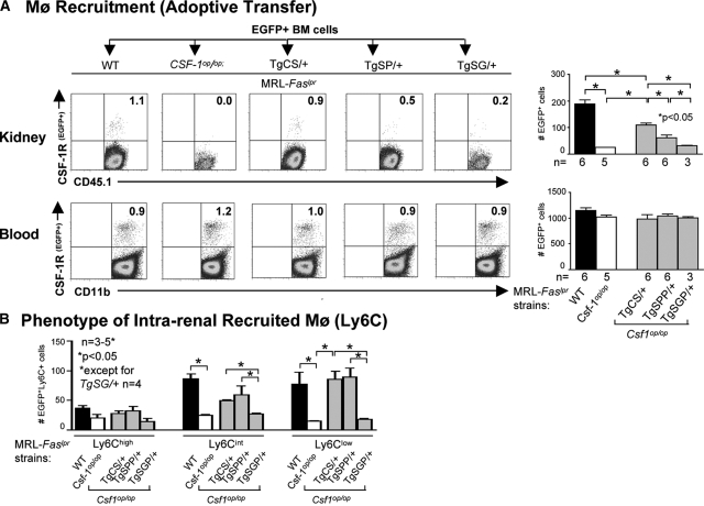
Figure 5.
Intrarenal csCSF-1 and spCSF-1 mediate the recruitment of activated monocytes into the kidney during lupus nephritis. (A) BM cells from MacGreen(eGFP+);MRL-Faslpr mice were transferred into TgCS/+, TgSPP/+, TgSG/+, wild-type (WT), and Csf1op/op; MRL-Faslpr mice and the recruitment of eGFP+ cells into the kidney 24 hours after transfer of cells were evaluated by flow cytometry. WT and Csf1op/op mice served as positive and negative controls, respectively. The data are representative of three experiments. The mice were 5 months of age. The values are the means ± SEM. Representative FACS plots are displayed. (B) Flow cytometric analysis of the recruited eGFP+ Mø for there Ly6Chi, Ly6Ciint, and Ly6Clo expression in kidney. The mice were 5 months of age. The values are the means ± SEM.

