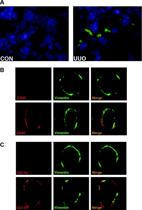
Figure 2.
Bone marrow-derived fibroblast precursors accumulate in the kidney in response to obstructive injury. (A) Representative photomicrographs show GFP-positive cells in the kidney in response to obstructive injury. Cryosections of the kidneys were fixed and stained with DAPI and examined with a fluorescence microscope. GFP, green; DAPI, blue. (B) Representative photomicrographs show that CD45- and vimentin-positive fibroblast precursors are present in the kidney in response to obstructive injury. Frozen kidney sections were stained for CD45 (red) and vimentin (green) and were examined with a confocal microscope (original magnification, ×1000). Upper panel shows representative photomicrographs of CD45 and vimentin staining of the control kidney. Lower panel shows representative photomicrographs of CD45 and vimentin staining of the obstructed kidney. CD45 and vimentin dual-positive fibroblast precursors are observed only in the kidney with obstructive injury. (C) Representative photomicrographs show that CD11b- and vimentin-positive fibroblast precursors are present in the kidney in response to obstructive injury. Frozen kidney sections were stained for CD11b (red) and collagen I (green) and were examined with a confocal microscope (original magnification, ×1000). Upper panel shows representative photomicrographs of CD11b and vimentin staining of the control kidney. Lower panel shows representative photomicrographs of CD11b and vimentin staining of the obstructed kidney. CD11b and vimentin dual-positive fibroblast precursors are observed only in the kidney with obstructive injury.

