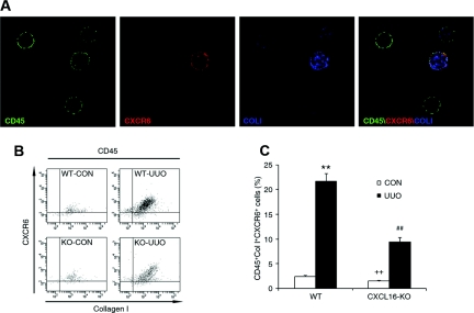
Figure 5.
Bone marrow-derived fibroblast precursors express functional CXCR6. (A) Bone marrow-derived fibroblast precursors express CXCR6. Peripheral blood cells were stained for CD45 (green), CXCR6 (red), and collagen I (blue) and identified by a deconvolution fluorescence microscope (original magnification, ×600). CD45, CXCR6, and collagen I triple-positive fibroblast precursors were detected in the circulation. (B) Representative cytometric diagrams showing the CXCR6- and collagen-I-positive cell distribution of all CD45-positive cells. Freshly isolated cells from the whole kidney of WT and CXCL16-KO mice with or without UUO for 5 days were stained with FITC-conjugated anti-CD45 antibody, PE-conjugated anti-CXCR6 antibody, and biotin-conjugated anti-collagen I antibody followed by APC-conjugated streptavidin; fluorescence intensities were measured by flow cytometry. (C) Quantitative analysis of CD45-, CXCR6-, and collagen-I-positive fibroblast precursors in the kidney of WT and CXCL16-KO mice with or without UUO as determined by flow cytometry. **P < 0.01 versus WT control, ++P < 0.01 versus KO UUO, and ##P < 0.01 versus WT UUO. n = 4 per group.

