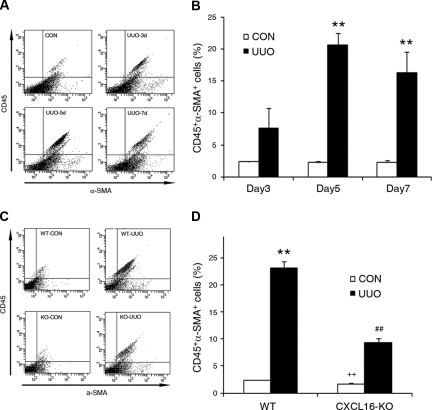
Figure 6.
CD45 and α-SMA dual-positive myofibroblasts accumulate in the injured kidney in a time- and CXCL16-dependent manner. (A) Representative cytometric diagrams showing CD45- and α-SMA-positive cells in the kidneys of WT mice with or without UUO at day 3, 5, and 7. Freshly isolated cells from the whole kidney of WT mice with or without UUO for 3, 5, or 7 days were stained with FITC-conjugated anti-CD45 antibody and PE-conjugated anti-α-SMA antibody; fluorescence intensities were measured by flow cytometry. (B) Quantitative analysis of CD45- and α-SMA-positive cells in the kidney of WT mice with or without UUO at day 3, 5, and 7 as determined by flow cytometry. **P < 0.01. n = 3 per group. (C) Representative cytometric diagrams showing CD45- and α-SMA-positive cell distribution in the kidney of WT and CXCL16-KO mice with or without UUO. Freshly isolated cells from whole kidney of WT and CXCL16-KO mice with or without UUO for 5 days were stained with FITC-conjugated anti-CD45 antibody and PE-conjugated anti-α-SMA antibody; fluorescence intensities were measured by flow cytometry. (D) Quantitative analysis of CD45- and α-SMA-positive cells in the kidney of WT and CXCL16-KO mice with or without UUO as determined by flow cytometry. **P < 0.01 versus WT control, ++P < 0.01 versus KO UUO, and ##P < 0.01 versus WT UUO. n = 4 per group.

