Fig. 3.
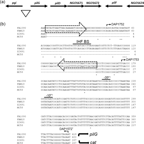
The pilGD locus. (a) Schematic representation of pilGD in N. gonorrhoeae strain FA1090. The triangle marks the region of DNA sequence examined. (b) Sequence alignment of the promoter regions for pilG in N. gonorrhoeae strains FA1090 and N. meningitidis strains Z2491, MC58, and FAM18. The dashed arrows enclose the two terminal CR. A −10 box and the corresponding −35 box of a σ70 promoter, termed P1, are shared by all strains and are overlined. The IHF BS contained within the CREE is overlined. Base differences among the four sequences are marked with asterisks. The curved solid arrows indicate the directions of pilG and cat transcription. The numbered arrows indicate the oligonucleotide primers used (see Table S2 in the supplemental material).

