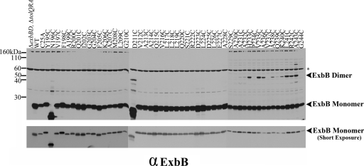
Fig. 7.
Cytoplasmic ExbB cysteine substitutions form disulfide-linked homodimers. The parent strain for all of the plasmid variants RA1017 (ΔexbBD ΔtolQRA), wild-type W3110 (WT), the parent plasmid encoding both ExbB and ExbD (pExbBD), and the plasmids expressing the ExbB variants are indicated above each lane of the immunoblot. To achieve chromosomal levels, the plasmid-encoded ExbB Y195C, N196C, D211C, Q215C, D223C, L224C, A228C, and G244C mutants were induced in tryptone broth with 0.00002, 0.005, 0.005, 0.00002, 0.00002, 0.00004, 0.00001, and 0.0001% l-arabinose, respectively. All other mutants were grown in tryptone broth without inducer. Samples were resolved on 13% nonreducing polyacrylamide gels and immunoblotted with anti-ExbB antibody. The figure is a composite of three immunoblots. The positions of the ExbB disulfide-linked dimers and ExbB monomer are indicated on the right. The positions of the mass markers are indicated on the left. The asterisk indicates the nonspecific cross-reactive bands. A shorter exposure of the immunoblot to allow comparison of monomer levels is presented in the lower panel.

