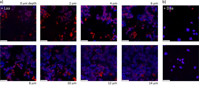
Fig. 5.
Confocal laser scanning microscopy images show no difference in the localization of EPS in S. aureus aggregates from cultures grown with d- or l-amino acids. These cells were stained with EPS-specific (blue) and DNA-specific (red) dyes, and the imaging sections were obtained in the same manner as those in Fig. 4. (a) Cultures treated with l-amino acids exhibited robust biofilms with localization of EPS around the cells in the aggregates. (b) Those treated with d-amino acids retained the EPS surrounding the cells but adhered as a submonolayer film, lacking the thickness and structure of a mature biofilm. As in Fig. 4c, the cells shown in panel b were attached only at the surface of the substrate. The scale bars are 5 μm.

