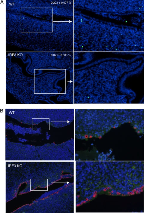
Fig. 4.
Reduced numbers of TUNEL-positive cells in the uterine horns but increased chlamydial infection in IRF3 KO mice in comparison to WT mice at day 4 postinfection. WT control (n = 5) and IRF3 KO (n = 5) mice were infected with 1 × 10⁶ IFU/ml of C. muridarum. At day 4 postinfection, genital tracts were excised and processed for TUNEL as described in Materials and Methods. (A) Images acquired using a 10× objective and a magnified section from a representative horn from each group are shown. Blue indicates DAPI-positive cells, and green indicates nuclear DNA fragmentation by TUNEL staining. Numbers on the panel indicate the percentages of TUNEL-positive cells in the uterine horns as determined from 10 images for each mouse (5 mice per group ± standard error of the mean) (P = 0.019, unpaired t test.) (B) Staining for chlamydial antigen in adjacent sections shows inclusions in epithelial cells. Blue indicates DAPI-positive cells, and red indicates chlamydial staining with antichlamydial mouse serum followed by Texas Red-conjugated anti-mouse antibody. A representative section is shown from each group at two magnifications (×100 [left] and ×400 [right]).

