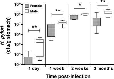
Fig. 2.
H. pylori colonization levels in male mice were elevated compared with those in females as early as 1 day postinfection. Male or female 129/Sv mice were infected with 107 H. pylori, and gastric CFU were assessed 1 day (n = 8), 1 week (n = 8), 2 weeks (n = 5), and 3 months (n = 8) later. Box plots present the median (horizontal bar), interquartile range (box), and 10th and 90th percentile ranges (whiskers). *, P < 0.03; **, P < 0.008 (ANOVA).

