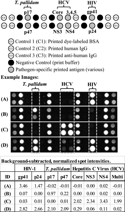
Fig. 2.
Representative images and array layout for the multiplex HIV/syphilis/HCV assay. The 30-feature array (2 rows by 15 columns) includes pathogen-specific printed antigens as well as multiple in-assay controls. Dye-labeled BSA spots serve as corner markers (C1) for imaging. These spots are not used in the analysis. Printed human IgG (C2) serves as a control for the dye-labeled detect antibody. Printed anti-human IgG (C3) serves as a sample control. Print buffer spots serve as a nonspecific binding negative control. Images from four clinical plasma samples are shown, along with background-subtracted spot intensities for the pathogen-specific antigens. Based on reference test methods, samples A to C are each monoinfected, with reactivity to HIV Ab (A), T. pallidum Ab (B), and HCV Ab (C). Sample D has Ab reactivity to all three pathogens by both the reference methods and the MBio assay. See the text for additional details.

