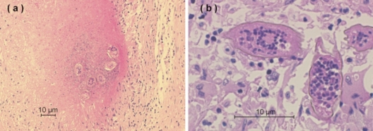
Fig. 2.
(a) Photomicrograph showing nodular granulomas within the parenchyma of the brain containing deposits of S. haematobium ova in the center of the granulomas (hematoxylin-eosin stained; magnification, ×100). (b) Ova of S. haematobium with a characteristic prominent terminal spine (hematoxylin-eosin stained; magnification, ×400).

