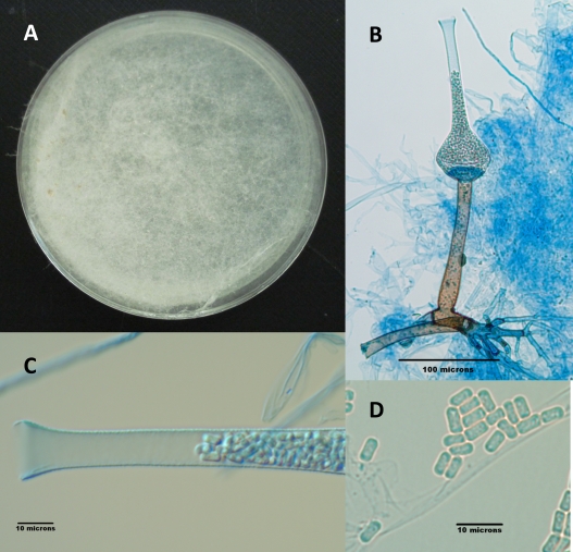
Fig. 2.
(A) S. erythrospora on CZA after 3 weeks of incubation at 25°C. (B) Fruiting structure of S. erythrospora depicting lateral rhizoids; a brown, straight, encrusted sporangiophore; the columella at the base of the sporangium; and the sporangium with its long neck filled with sporangiospores. (C) Higher magnification of the neck of the sporangium filled with sporangiospores. (D) Ellipsoidal, biconcave sporangiospores.

