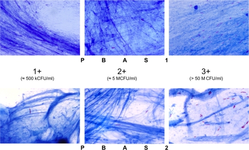
Fig. 3.
Comparative microscopic appearances showing the different positivity grades. An image for the ± grade is not shown, because it cannot be distinguished from the + grade in a pair of single pictures. In PBAS2, bacilli are stained dark and are more clearly distinguished than in the PBAS1 smears.

