Fig. 6.
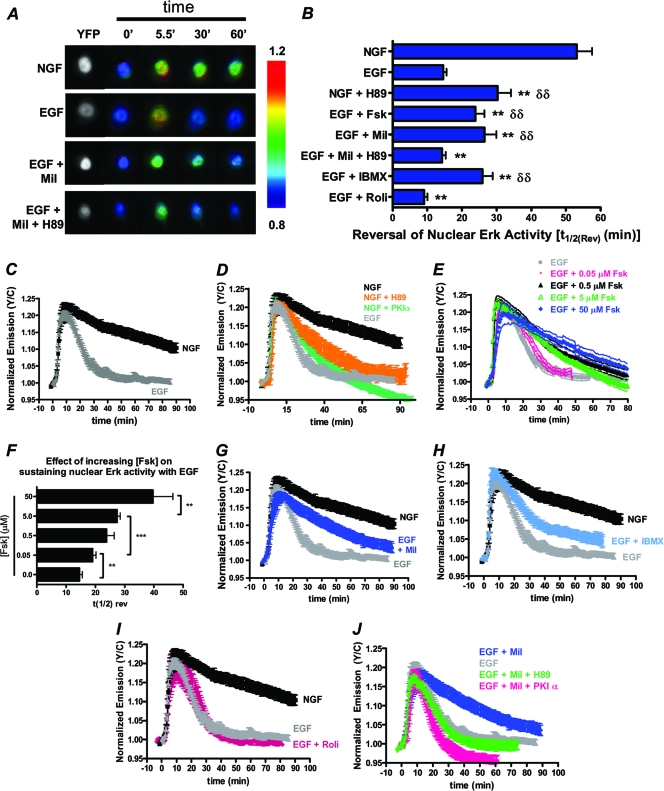
PDE3 inhibition extends the duration of EGF-induced nuclear ERK in a PKA-dependent manner. All experiments are done in PC12 cells expressing EKARnuclear. (A) YFP direct (left) and ratiometric images depicting differential GF-induced nuclear ERK activities in the presence and absence of PDE3 inhibitor (Mil) and/or PKA inhibitor (H89). (B) Graphical representation of the time needed for nuclear ERK activity to reduce to 50% [t1/2(Rev)]. (C) NGF-induced nuclear ERK activity (n = 9) is more sustained than EGF-induced nuclear ERK activity (n = 10). (D) Time course of NGF-induced nuclear ERK activity in the presence of H89 (n = 8) and PKIα (n = 5) expression. (E) tmAC activation with increasing doses of Fsk extends the duration of EGF-stimulated ERK activity in the nucleus (0.05 μM Fsk, n = 8; 0.5 μM Fsk, n = 9; 5.0 μM Fsk, n = 10; 50 μM Fsk, n = 7. (F) Bar graph depicting the increase in reversal of nuclear ERK activity [t1/2(Rev)] following various treatments (EGF plus the following doses of Fsk: 0.05 μM, n = 16; 0.5 μM, n = 17; 5.0 μM, n = 10; 50 μM Fsk, n = 7). (G) PDE3 inhibition with 10 μM Mil (n = 7) extends the duration of EGF-stimulated ERK activity. (H and I) 100 μM IBMX increases the duration of EGF-stimulated nuclear ERK activity (n = 8) (H), while 1 μM Roli does not (n = 7) (I). (J) In the presence of milrinone and inhibition of PKA through H89 pretreatment (n = 8) or PKIα expression (n = 5), there is no increase in the duration of EGF-induced nuclear ERK activity. All data are shown as means ± SEMs. δδ, P < 0.01 compared to EGF. **, P ≤ 0.01 compared to NGF.

