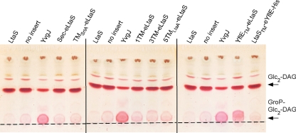
Fig. 6.
TLC analysis of glycolipids produced by S. aureus strains expressing different LtaS fusion proteins. The LtaS fusion protein indicated above each lane was expressed from a multicopy plasmid by the addition of Atet after removal of IPTG in the S. aureus LtaS depletion strain ANG499. Strains expressing S. aureus LtaS and B. subtilis YvgJ and a strain containing the empty plasmid pCN34 (no insert) were used as controls. Cultures were grown to mid-log phase, and lipids were extracted and separated by TLC as described in Materials and Methods. Glycolipids were visualized by staining with α-naphthol and sulfuric acid. The origin is marked with a dashed line, and the positions of the glycolipids Glc2-DAG (top band) and GroP-Glc2-DAG (bottom band) are indicated with arrows on the right. S. aureus strains ANG1130, ANG1571, ANG1658, and ANG2165 to ANG2171 were used for this experiment.

