
Fig. 1.
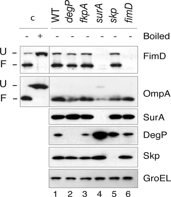
FimD folding in E. coli null mutants in periplasmic chaperones. Western blot analysis of whole-cell protein extracts from E. coli UT5600 (WT) strain and degP, fkpA, surA, skp, and fimD null mutants developed with anti-FimD, anti-OmpA, anti-SurA, anti-DegP, and anti-Skp antibodies, as indicated on the right of each panel. Detection of cytoplasmic GroEL with specific antibodies is used as a loading control (lower panel). Whole-cell extracts were prepared in SDS sample buffer and not boiled for detection of the folded forms of FimD and OmpA. Control sample c+ was boiled in urea-SDS sample buffer before loading in order to fully denature FimD and OmpA. The mobility of the protein bands corresponding to folded (F) and unfolded (U) FimD and OmpA are labeled on the left. Control samples are obtained from a culture of the WT strain.